HQ Team
September 24, 2025: Scientists at the University of Technology Sydney (UTS) have built a highly accurate model of the early human placenta using 3D bioprinting. This will help scientist overcome a critical limitation as the present studies rely on animal or conventional cell models that do not accurately present human placental biology.
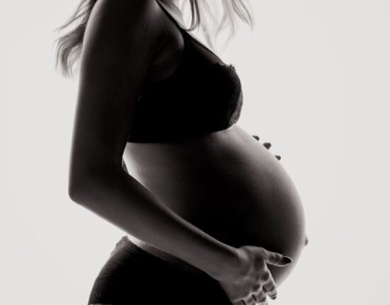
3D bioprinted mini placentas represent a major breakthrough in understanding early pregnancy and associated complications like preeclampsia, which affects 5–8% of pregnancies and contributes to over 260,000 maternal and millions of infant deaths globally.
Placental research challenges
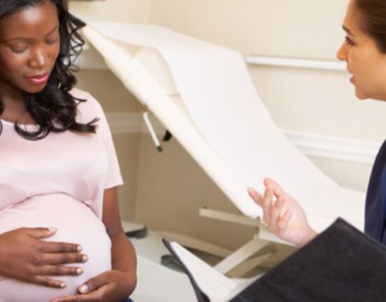
Studying early human placental tissue is extremely difficult because obtaining first trimester samples is neither practical nor safe. By the time of birth, placental tissue undergoes significant changes, making it unrepresentative of its early pregnancy form. This gap in accessible, reliable models hampers understanding of placental dysfunctions that underlie serious pregnancy complications such as preeclampsia, a condition still poorly understood despite its high impact on maternal and infant health .
Early advances in organoid technology provided a foundation for placental research by allowing the growth of mini organs (organoids) from stem cells in a supporting gel that simulates the native tissue environment. Placental organoids were first grown in 2018 using trophoblast cells, which are exclusive to the placenta. These organoids served as a breakthrough over traditional cell cultures but still had limitations due to the animal-derived gels traditionally used, which might alter how cells develop and mature .
“Obtaining first trimester placental tissue is not practical or safe, making early pregnancy challenging to study. By the time a baby is born, the placenta has changed so much that it no longer reflects what it was like in early pregnancy,” said Dr Lana McClements, the lead of the study.
The 3D bioprinted mini placentas
The UTS team introduced 3D bioprinting, a technique using living cells combined with synthetic gels that can be precisely manipulated. They mixed trophoblast cells with a controllable synthetic gel and printed them into culture dishes in fine droplets, much like inkjet printing. This method allowed better control of the cellular microenvironment, leading to organoids that developed differently and more closely resembled early human placental tissue compared to those grown in animal-derived gels .
“Our printed cells grew into placental organoids and we compared them to organoids made via traditional manual methods,” said Clara Dr Richards, co-author of the study.
“We showed these organoids were very similar to human placental tissue, providing an accurate model of the early placenta. This means we can start piecing together the puzzle of pregnancy complications and test new drugs safely.
“For example, we exposed our bioprinted organoids to an inflammatory molecule found at high levels in women with preeclampsia, then tested potential treatments to see how the organoids grew and responded.
“As we refine these models, we move closer to a future where pregnancy complications can be predicted, prevented and treated before they put lives at risk.”
Their study demonstrated that the bioprinted mini placentas contain diverse sub-types of trophoblast cells and behave in ways very similar to natural early placental tissue. This authenticity enables a more reliable model for studying how the placenta forms and functions during early pregnancy, which is the period when many complications originate .
Pregnancy research and future benefits
The ability to bioprint accurate mini placentas opens new avenues for investigating pregnancy complications like preeclampsia at the cellular and molecular levels. Researchers used inflammatory molecules present in women with preeclampsia to test how these organoids respond and react to potential treatments. This approach allows safe, ethical, and detailed study of disease mechanisms and drug testing without risking human or animal subjects .
Going forward, this technology could enable prediction, prevention, and personalized treatment of pregnancy complications before they cause harm, potentially saving thousands of lives annually. It also sets a precedent for using bioprinted organoids to model other complex human tissues where research is limited by tissue access and ethical concerns .
Organoid technology was introduced in 2008 and revolutionized tissue modeling. The first placental organoids were grown from trophoblasts in animal-derived gels in 2018.
This study directly addresses challenges in studying early human placental development and disease, which previous models failed to capture adequately .It promises to accelerate discoveries into the causes and treatments of pregnancy complications and improve maternal and infant health outcomes worldwide.
The study, led by Associate Professor Lana McClements and first author Dr Claire Richards, from the UTS School of Life Sciences, has just been published in the journal Nature Communications.