HQ Team
October 2, 2025: University of Cambridge scientists have developed a first-time imaging technique that helps visualise and quantify the early triggers of Parkinson’s — the world’s fastest-growing neurological disease.
The tiny protein cluster, called alpha-synuclein oligomers, has long been considered the likely culprit for Parkinson’s disease to start developing in the brain, but until now, the proteins have evaded direct detection in human brain tissue.
The imaging method allows them to see, count and compare oligomers in human brain tissue. It is “like being able to see stars in broad daylight,” the researchers said, according to a statement from the university.
For more than a century, doctors have recognised Parkinson’s by the presence of large protein deposits called Lewy bodies. Scientists have suspected that smaller, earlier-forming oligomers may cause damage to brain cells.
Until now, these oligomers were simply too small to see – just a few nanometres long.
Oligomers
Researchers from the University of Cambridge, the Francis Crick Institute and Polytechnique Montréal said the discovery could help unravel the mechanics of how Parkinson’s spreads through the brain and support the development of diagnostics and potential treatments.
Alpha-synuclein oligomers are small clumps made up of several alpha-synuclein proteins stuck together inside brain cells.
Normally, alpha-synuclein helps brain cells communicate by controlling the release of chemicals called neurotransmitters. But sometimes, these proteins start sticking to each other abnormally, forming these clumps called oligomers.
These oligomers are important because they are thought to be harmful to brain cells. They can damage cell membranes and other parts of the cell, which may lead to brain cell death. This process is linked to diseases like Parkinson’s disease, where these harmful protein clumps build up and cause problems with movement and brain function.
Alpha-synuclein oligomers come before even larger clumps called fibrils or Lewy bodies, which are seen in affected brains. Stopping these oligomers from forming or reducing their toxicity can help in finding treatments.
25 million by 2025
About 166,000 people in the UK live with Parkinson’s disease, and the number is rising. By 2050, the number of people with Parkinson’s worldwide is expected to double to 25 million.
While there are drugs that can help alleviate some of the symptoms of Parkinson’s, such as tremor and stiffness, there are no drugs that can slow or stop the disease itself.
“Lewy bodies are the hallmark of Parkinson’s, but they essentially tell you where the disease has been, not where it is right now,” said Professor Steven Lee from Cambridge’s Yusuf Hamied Department of Chemistry, who co-led the research.
“If we can observe Parkinson’s at its earliest stages, that would tell us a whole lot more about how the disease develops in the brain and how we might be able to treat it.”
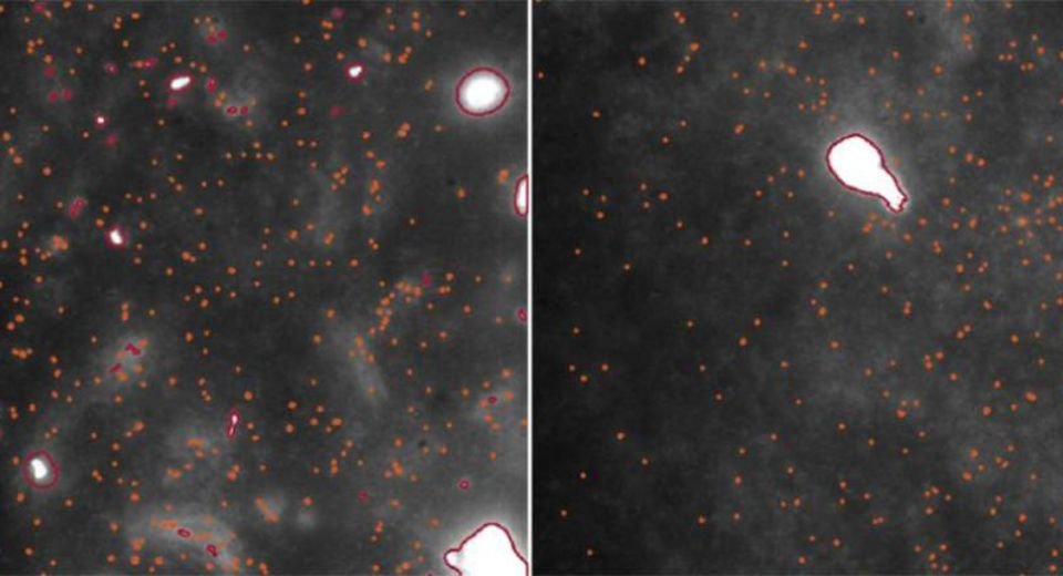
The imaging technique uses ultra-sensitive fluorescence microscopy to detect and analyse millions of oligomers in post-mortem brain tissue. Since oligomers are so small, their signal is extremely weak.
Post-mortem brain tissue samples
The technique maximises the signal while decreasing the background, dramatically boosting sensitivity to the point where individual alpha-synuclein oligomers can be observed and studied.
“This is the first time we’ve been able to look at oligomers directly in human brain tissue at this scale: it’s like being able to see stars in broad daylight,” said co-first author Dr Rebecca Andrews, who conducted the work when she was a postdoctoral researcher in Lee’s lab. “It opens new doors in Parkinson’s research.”
The team examined post-mortem brain tissue samples from people with Parkinson’s and compared them to healthy individuals of similar age.
They found that oligomers exist in both healthy and Parkinson’s brains. The main difference between disease and healthy brains was the size of the oligomers, which were larger, brighter and more numerous in disease samples, suggesting a direct link to the progression of Parkinson’s.
The team also discovered a subclass of oligomers that appeared only in Parkinson’s patients, which could be the earliest visible markers of the disease, potentially years before symptoms appear.
“This method doesn’t just give us a snapshot,” said Professor Lucien Weiss from Polytechnique Montréal, who co-led the research. “It offers a whole atlas of protein changes across the brain, and similar technologies could be applied to other neurodegenerative diseases like Alzheimer’s and Huntington’s.
“Oligomers have been the needle in the haystack, but now that we know where those needles are, it could help us target specific cell types in certain regions of the brain,” he said.