Bharti Jayshankar
January 24, 2024: Researchers in the UK and Switzerland have successfully grown miniature placentas in a laboratory setting, offering new insights into the origins of pregnancy disorders such as pre-eclampsia, stillbirth, and growth restrictions. The development of these “mini-placentas” provides a crucial window into the intricate processes occurring in the first weeks of pregnancy, where disorders can take root and potentially harm both the mother and the developing baby.
Decoding the human black box
Successful pregnancy hinges on the proper development of the placenta during the initial weeks of gestation. This critical phase sees the placenta embedding itself into the endometrium, the mucosal lining of the uterus. Unfortunately, disorders like pre-eclampsia often emerge during this enigmatic period, making it challenging for scientists to study and comprehend.
Pre-eclampsia
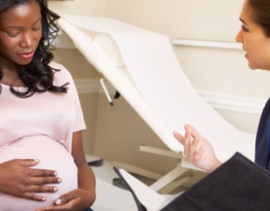
Pre-eclampsia, a condition affecting about 6 in 100 first pregnancies, arises when interactions between endometrial and placental cells go awry. This disorder, characterized by high blood pressure during pregnancy, poses significant risks to both the expectant mother and the unborn child. The lack of understanding about the intricate dynamics during implantation makes studying and preventing such disorders a formidable task.
Mini-placentas
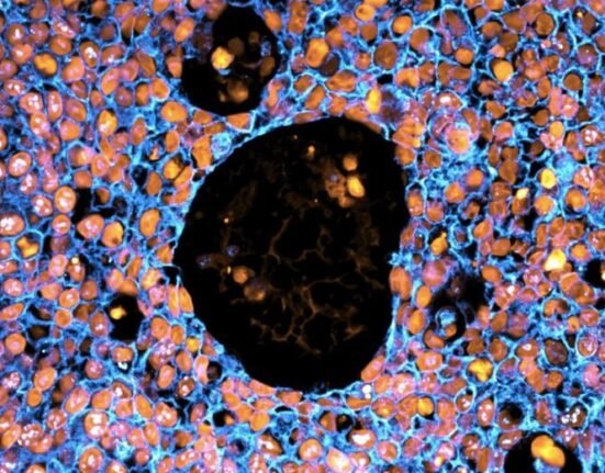
To address the complexities of early pregnancy development, scientists, including Professor Ashley Moffett from the University of Cambridge, experimented with mini-placentas, or “trophoblast organoids.” These lab-grown structures allowed researchers to delve into the interactions between proteins extracted from immune cells in the uterus and the placenta during implantation.
Key role of proteins
The study, detailed in Cell Stem Cell, revealed that proteins from uterine immune cells play a pivotal role in the success of placental implantation. Professor Moffett emphasized that this phase, where normal cells from the baby transform an artery, is unique. Proper invasion ensures the opening of arteries in the womb, supplying nutrients and oxygen to the placenta and the growing fetus. Failure in this process can result in severe complications later in pregnancy.
A new perspective on pre-eclampsia
Margherita Turco from the Friedrich Miescher Institute, who co-led the study with Professor Moffett, highlighted the persistent lack of understanding surrounding pre-eclampsia. By utilizing mini-placentas, researchers aim to unravel crucial processes in the early weeks of pregnancy, offering a new approach to predict and prevent this condition. Turco emphasized the potential of basic science to enhance our comprehension of fundamental biology, paving the way for significant advancements in maternal and infant health.
Turco underscored the potential impact of basic science in deciphering fundamental biological processes. The hope is that these discoveries will contribute to future breakthroughs, ultimately making a substantial difference in safeguarding the health of both mothers and their newborns.