HQ Team
July 1, 2025: Medical experts at Cincinnati Children’s have made an organoid, or ‘organs in a dish,’ of human lung tissue that produces its own blood vessels, according to a statement.
Before “our study, the development of lung organoids with organotypic vasculature had not been achieved,” says co-corresponding author Mingxia Gu, MD, PhD. “Notably, this method also could be applied to other organ systems such as the intestine and colon.”
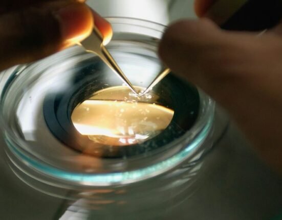
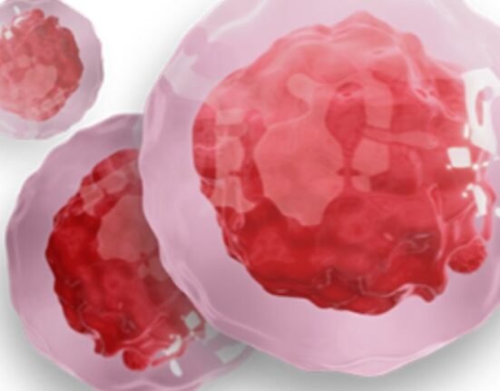
Creating organoids involves converting mature human cells, such as blood or tissue cells, into fetal-like stem cells that can be coaxed to grow a wide range of other tissue types.
Unlike disconnected human cells kept alive in a dish, these are growing, developing mini-organs that form into seed-sized spheres which mimic the unique functions of full-sized organs.
Examples include the intestines that absorb and secrete. Stomachs that produce digestive acids. Hearts that pulse and brain tissues that fire nerve cells.
Integration
Cincinnati Children’s has been a leader in organoid development since 2010 when experts here produced the world’s first functional intestinal organoid grown from induced pluripotent stem cells (iPSCs).
The challenge has been learning how to grow organoid tissues that can connect with the rest of the body to integrate nerve connections, blood vessels, bile ducts, immune systems and more.
During pregnancy, these differing tissue types naturally find each other as the fetus matures and becomes more complex.
Organoid developers are seeking to reproduce these steps in the laboratory, which eventually may allow people to receive custom-grown tissues that could patch damage or boost disrupted functions.
Simpler forms of organoids have already begun transforming medical research, allowing many scientists to use living human tissue models to study disease while reducing current reliance on animal models to develop new medicines.
Four years of research
Without the ability to make internal blood vessels, the tiny seeds lack the ability to grow into larger, more useful tissues.
The Cincinnati study spanned four years and involved more than 20 people at Cincinnati Children’s and collaborations with experts at several other organisations.
Co-first and co-corresponding author Yifei Miao, PhD, now at the Chinese Academy of Sciences, Beijing, was with Cincinnati Children’s for this work.
“The challenge in vascularizing endodermal organs, particularly the lung, stems from different signalling requirements for lung epithelial versus vascular differentiation,” Miao said. “Our success in this endeavour is attributable to our unique differentiation method.”
The team grew iPSCs from multiple cell types, then found the right moment to introduce them to each other. The resulting cell signals helped flip a developmental switch so that progenitor cells that could have become either blood vessels or the outer walls of the lung wound up becoming blood vessels.
Lungs’ air sacs
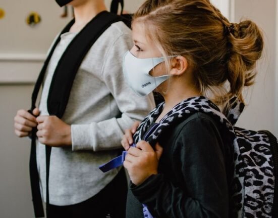
Advanced lab-grown tissues help show how special lung cells develop, shedding light on rare Alveolar Capillary Dysplasia with Misalignment of Pulmonary Veins disease and suggesting potential ways to repair damage from viral infections such as Covid-19.
Within days of birth, infants born with ACDMPV struggle to breathe because their lungs’ air sacs (alveoli) and blood vessels are malformed. Nearly all infants with this condition die within the first month of life.
Cincinnati Children’s has filed patent applications related to the methods developed here to produce organoids with blood vessel formation capabilities, and the team is moving to further develop this technology.
“We look forward to continuing to learn more about the fundamental biology involved in organ formation and applying those discoveries to improving outcomes across a wide range of difficult human diseases and conditions,” said Aaron Zorn, PhD, co-director of the Center for Stem Cell and Organoid Medicine at Cincinnati Children’s team and director of the Division of Developmental Biology.